Cellular Bioactivity: The Ultimate Functional Assay to Decode Active Protein Function
Cell-based functional testing delivers physiologically-relevant readouts for recombinant protein characterization
Press Release Disclaimer: This is a press release distributed through the XPR Media network. It has not been independently verified by our newsroom.

Cell-based functional testing delivers physiologically-relevant readouts for recombinant protein characterization across research and biomanufacturing
HUSTON, TX, UNITED STATES, October 29, 2026 /EINPresswire.com/ — Recombinant active proteins serve as indispensable tools for basic life-science research, cell-therapy manufacturing and biopharmaceutical development. While molecular-level binding assays and enzymatic activity measurements provide valuable molecular evidence, they cannot fully replicate complex physiological responses within living cells. Cell-based bioactivity assays represent the gold-standard functional readout, directly demonstrating whether a protein can trigger expected cellular behaviours. Cloud-Clone outlines core concepts, common assay formats, decision-making criteria and industry-wide applications for cellular bioactivity evaluation to support global researchers and biotech developers.
Cellular bioactivity acts as the ultimate assessment benchmark for validating active protein function. To verify swimming capability, direct in-water testing delivers definitive proof. Similarly, the most physiologically-relevant approach for confirming the true biological function of an active protein is observing its ability to elicit responses in living cells.
Among three major validation systems for active proteins, cellular bioactivity testing best mimics native in-vivo physiological conditions. Binding assays confirm target-molecule recognition, and enzyme-activity assays verify catalytic performance. Cellular bioactivity testing answers a critical downstream question: can this protein trigger anticipated biological effects inside living cells? Observable, quantifiable cellular outcomes including proliferation, migration, differentiation and apoptosis serve as conclusive functional evidence for active proteins.
For this reason, cell-based bioactivity assays constitute mandatory release testing for many biopharmaceutical products. For functionally-regulating proteins such as cytokines, growth factors and chemokines, cellular bioactivity defines product performance.
What is cellular bioactivity?
Cellular bioactivity describes the capacity of an active protein to induce specific morphological, numerical, behavioural or physiological changes within living-cell systems. Proteins bind to cell-surface targets, activate intracellular signalling cascades, modulate gene expression, and ultimately generate measurable cellular responses. Cellular bioactivity represents the integrated output of this complete signalling chain.
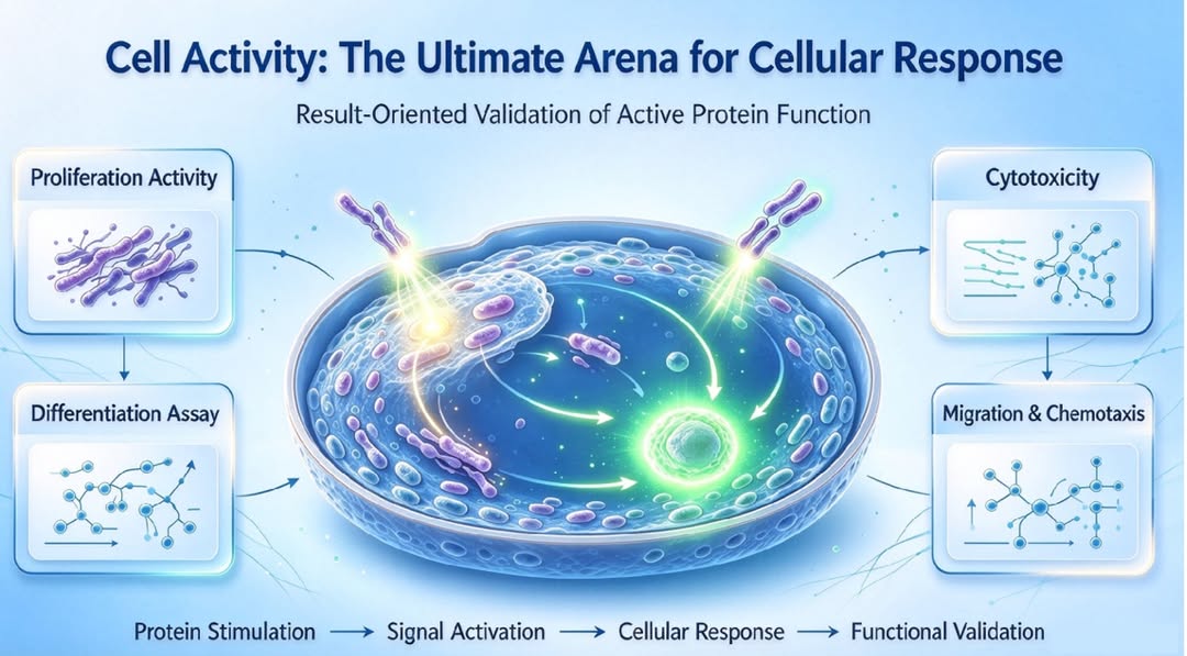
Distinct from molecular-scale binding-activity tests, cellular bioactivity assays operate on intact living cells and capture complex biological reactions. Upon protein stimulation, cells integrate multiple incoming signals and produce final measurable responses, including:
Cell proliferation: Protein stimulation drives cell division and increases viable-cell counts
Growth inhibition / cell death: Suppressed cell proliferation or induction of apoptosis and necrosis
Cellular differentiation: Direct transformation of stem or progenitor cells into specialized functional cell types
Directed migration: Chemotactic signals guide directional cellular movement
Functional activation: Immune-cell stimulation or modified secretion profiles
These quantifiable cellular behaviours triggered by protein exposure provide key metrics for evaluating biological function. Cell-based bioactivity assays reconstruct near-physiological protein functions and form an irreplaceable component within active-protein validation workflows.
Figure 1 Cellular bioactivity: the ultimate assessment benchmark for cellular responses. Outcome-oriented validation for active-protein function verification. Protein stimulation → signalling activation → cellular response → functional confirmation)
Primary categories of cellular bioactivity assays
Based on testing endpoints, major cell-based functional assays fall into four key groups:
Proliferation Assays These assays verify whether a protein stimulates cell division and increases viable-cell populations, forming the core functional evaluation for cytokines and growth factors. Common approaches include CCK-8 / MTT assays, which indirectly quantify viable-cell numbers via metabolic activity; EdU and BrdU incorporation assays, which directly label DNA synthesis to visualize proliferation intensity. Applicable proteins: Epidermal Growth Factor (EGF), basic Fibroblast Growth Factor (FGF2/bFGF), Insulin-like Growth Factor (IGF), Interleukin-2 (IL-2), and others.
Cytotoxicity / Growth-Inhibition Assays These measure a protein’s capacity to kill target cells or suppress cellular growth, representing core evaluation workflows for Tumour Necrosis Factor and functional therapeutic antibodies. Common approaches: LDH-release assays for detecting cell-membrane damage; Annexin V / PI dual-staining coupled with flow cytometry to distinguish apoptotic and necrotic cell populations. Applicable proteins: Tumour Necrosis Factor-α (TNF-α), selected anti-tumour functional antibodies.
Differentiation Assays These validate protein-driven directed differentiation of stem or progenitor cells toward specialized functional phenotypes. Common approaches: Microscopic morphological observation (such as neurite outgrowth for neuronal cells or mineralization for osteoblasts); flow cytometry or qPCR detection of unique differentiation biomarkers. Applicable proteins: Nerve Growth Factor (NGF), Bone Morphogenetic Proteins (BMPs), and related molecules.
Migration / Chemotaxis Assays These examine whether a protein can trigger directional cellular movement. The classic Transwell chemotaxis chamber setup divides the experimental system into upper and lower compartments by a microporous membrane. Researchers quantify cells migrating through pores along a protein concentration gradient. Applicable proteins: Chemokines including MCP-1, SDF-1, and other related factors.
Figure 2. Validation of recombinant human EGF‑driven proliferation in 3T3 fibroblasts.
How to determine whether cellular-bioactivity testing is required
Not every active protein mandates cell-based bioactivity testing. Requirements are defined by each protein’s biological functional profile:
Cytokines, growth factors, chemokines: Cellular bioactivity serves as an essential core metric, as their biological functions are manifested through modified cellular behaviours.
Hormonal proteins: Cellular bioactivity generally acts as a primary validation endpoint. For instance, the glucose-lowering effect of insulin must be validated at cellular or animal-model levels.
Antibodies: Binding affinity forms the fundamental baseline metric. Functional antibodies such as neutralizing antibodies and ADCC-capable antibodies require supplementary cell-based assays for full biological-effect characterization.
Enzymatic proteins: Enzymatic activity constitutes the primary quality-control marker; cellular-bioactivity testing is not routinely required for batch release.
For batch-release quality-control workflows, manufacturers select assays that best represent each protein’s core biological function as pass-fail criteria.
Industry-wide application scenarios for functionally-validated active proteins
Only recombinant proteins with stable, verified cellular bioactivity can exert genuine biological effects. Such materials are widely deployed across basic research, biopharmaceuticals, cell-therapy production and tissue-regeneration applications. Cellular bioactivity acts as the prerequisite for all these downstream use-cases.
1.Basic-research reagents Active proteins such as FGF2, EGF, IL-2, MCP-1, BMP and NGF serve as critical supplements for cell-culture systems. Researchers utilise these factors to induce proliferation, directional migration and stem-cell differentiation for building in-vitro cellular models for mechanistic investigations. Even with high purity and correct concentration, loss of cellular bioactivity will lead to complete experimental failure.
2.Recombinant-protein therapeutics Pharmacological efficacy of clinical recombinant-protein drugs relies on protein-triggered cellular responses. Examples include recombinant IL-2 for immune-cell proliferation and activation, BMP for osteogenic stem-cell differentiation, and EGF for wound repair. Pharmacopoeia guidelines designate cellular bioactivity as a mandatory release criterion for these medicines; batches failing bioactivity benchmarks cannot be released for clinical use.
3.Upstream raw materials for cell-therapy manufacturing During production of CAR-T, NK and mesenchymal stem cells, bioactive proteins including IL-2 and FGF2 are supplemented within culture media to expand immune-cell populations and sustain stem-cell pluripotency. Consistent cellular bioactivity directly impacts final viable-cell yield and product quality, making these materials critical upstream components for industrial-scale cell-therapy workflows.
4.Tissue repair and regenerative medicine Growth factors such as FGF2 and EGF exert proliferation- and migration-promoting effects for research and formulation development targeting skin, bone and nerve injury repair. Target-cell behaviour modulation enables tissue reconstruction and wound healing.
Cellular bioactivity bridges molecular-protein properties and physiological cellular responses. For both academic exploration and industrial translation, authentic bioactivity data, batch-to-batch consistency and full traceability underpin reliable experimental and clinical outcomes.
SuKi Duan
CLOUD-CLONE CORP.WUHAN
email us here
Visit us on social media:
LinkedIn
Instagram
TikTok
X
Legal Disclaimer:
EIN Presswire provides this news content “as is” without warranty of any kind. We do not accept any responsibility or liability
for the accuracy, content, images, videos, licenses, completeness, legality, or reliability of the information contained in this
article. If you have any complaints or copyright issues related to this article, kindly contact the author above.

Media gallery